 |
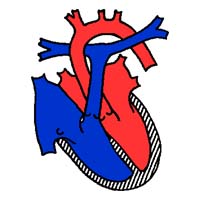
NORMAL HEART
 |
ALEXIS'S HEART
(Before Surgery)
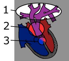 |
Alexis was diagnosed with
the following:
1) Interrupted Aortic Arch Type B
2) Truncus Arteriosis Type I
3) Ventricular Septal Defect
These are very big words for such a little heart.
1) Interrupted Aortic Arch Type B
- What is Interrupted Aortic Arch?
The aorta is the main blood vessel that carries oxygen-rich blood away from the heart to
the organs of the body. After it leaves the heart it ascends in the chest to give off
blood vessels to the arms and head, then arches to turn down toward the lower half of the
body. Interrupted Aortic Arch (IAA) is the absence or discontinuation of a portion of the
aortic arch.
What are the results of surgery?
The risk of complications both early and late following the repair of interrupted aortic
arch with VSD depends on a number of factors. Very small size of the aortic valve region
or significant instability in the preoperative period increase the chance of later
problems. Survival after complete repair of the aortic arch and VSD in the newborn period
is 90 percent or greater in most pediatric heart centers. Long term follow-up by the
cardiologist to assess growth of the aortic valve region and the reconstructed aortic arch
is essential. Reoperation to address further problems with these areas may be needed in 10
percent to 20 percent of patients.
-
- 2) Truncus Arteriosis Type I
-
- What is Truncus Arteriosus?
Normally there are two main blood vessels leaving the heart: the aorta carrying blood to
the body and the pulmonary artery that branches immediately to carry blood to each lung.
Instead of having a separate pulmonary artery and aorta, each with their own three-leafed
valves, a baby with truncus arteriosis has only one great blood vessel or trunk leaving
the heart, which then branches into blood vessels that go to the lungs and the body. This
great vessel usually has one large valve which may have between two and five leaflets.
Usually this great vessel sits over both the left and right ventricle. The upper portion
of the wall between these two chambers is missing resulting in what is known as a
ventricular septal defect, or VSD. In rare cases, the VSD is absent.
What are the results of surgery?
Currently over 90 percent of children survive repair of truncus Arteriosis. As
the child grows, they will be followed by their cardiologist. Typically there will be no
physical restrictions imposed on the child. As a child grows, the conduit that was used to
connect the right ventricle and pulmonary arteries will not, and this will lead to
obstruction to blood flow. The progression of this narrowing is easily followed by the
cardiologist using physical examination and echocardiograms. Recommendation for surgery to
replace the right ventricle to pulmonary artery conduit with a larger one is usually made
long before any symptoms would be evident. This conduit will often need to be replaced two
or three times during childhood to accommodate for growth. These operations are typically
tolerated very well with a hospitalization of less than a week.
3) Ventricular Septal Defect
- What is a Ventricular Septal Defect?
A ventricular septal defect, or VSD, is a hole between the right and left pumping chambers
of the heart. The heart has four chambers: a right and left upper chamber called an atrium
and a right and left lower chamber called a ventricle. In the normal heart the right and
left chambers are completely separated from each other by a wall called a septum. The
right atrium is separated from the left atrium by the atrial septum and the right
ventricle is separated from the left ventricle by the ventricular septum. It is normal for
all infants to be born with a small hole between the two atria, which usually closes
within the first few weeks of life. Normally there is no hole between the two ventricles,
but some infants are born with these holes called VSDs. It is one of the most common
congenital heart defects occurring in between 0.1 percent to 0.4 percent of all live
births and makes up about 20 percent to 30 percent of congenital heart lesions. It is
probably one of the most common reasons for referral of an infant to a cardiologist.
___________________________________________________________ |